
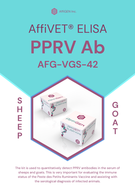

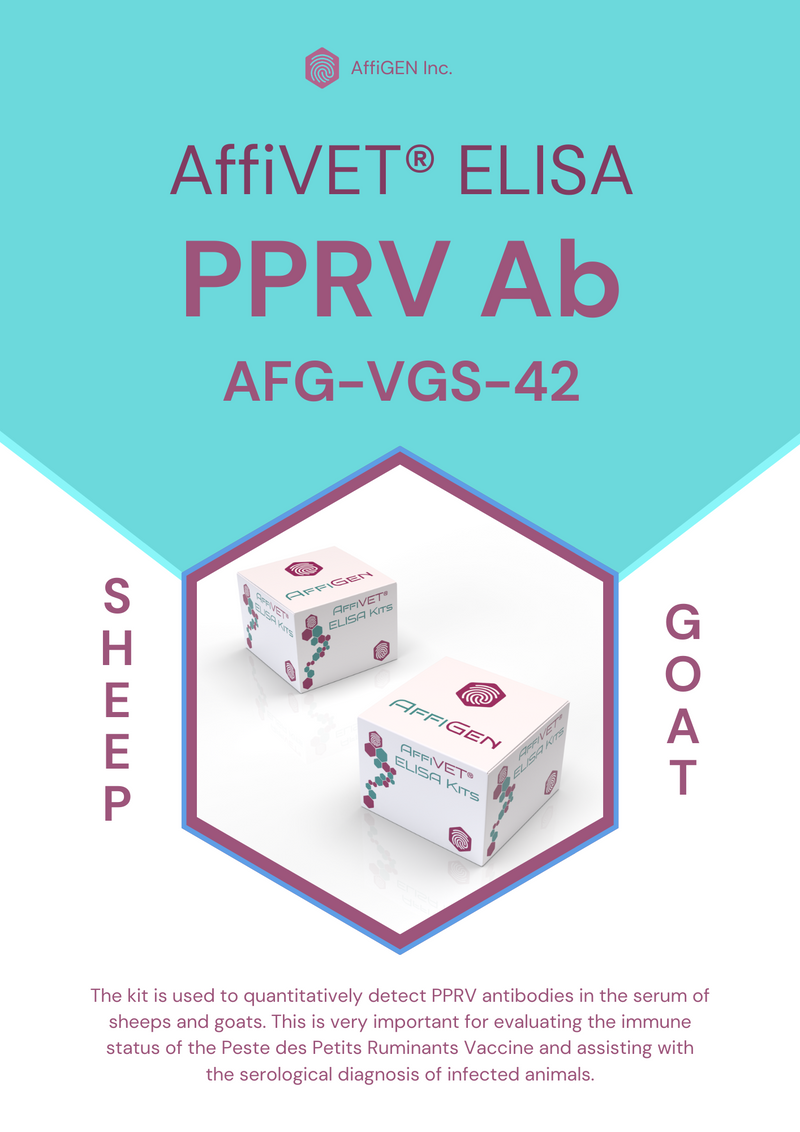


AffiVET® Peste des petits ruminants virus (PPRV) antibody ELISA kit
Cat Number: AFG-VGS-42
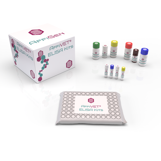
Size: 96 Well
Species: Sheeps & Goats
Sample: Serum
The kit is used to quantitatively detect PPRV antibodies in the serum of sheeps and goats. This is very important for evaluating the immune status of the Peste des Petits Ruminants Vaccine and assisting with the serological diagnosis of infected animals.
Overview:
- The kit uses competitive ELISA method with pre-coated PPRV antigen on enzyme micro-well strips.
- During testing, serum samples are mixed with Monoclonal Enzyme conjugate and incubated.
- If PPRV-specific antibodies are present in the sample, they bind to the PPRV antigen, blocking the enzyme-labeled monoclonal antibody from binding.
- Conversely, if no PPRV-specific antibodies are present, there is no binding to the coating plate.
- Unbound components are removed through washing.
- Substrate is added to the micro-wells, resulting in a blue product formation via enzymatic catalysis.
- The reaction is terminated with stop solution.
- The absorbance A value in the reaction well is measured using a microplate reader at 450 nm and 630 nm wavelengths.
Kit Components:
| PPRV Antigen Coated Microplate | 96 Tests | 2X 96 Tests | 96Tx4 | 5X 96 Tests |
| Enzyme Conjugate | 6 mL | 12 mL | 24ml | 30 mL |
| Sample Dilution | 6 mL | 12 mL | 24ml | 30 mL |
| Negative Control Serum | 0.75 mL | 1.5 mL | 3ml | 4 mL |
| Positive Control Serum | 0.75 mL | 1.5 mL | 3ml | 4 mL |
| Substrate | 12 mL | 2X 12 mL | 48ml | 60 mL |
| Stop Solution | 6 mL | 12 mL | 24ml | 30 mL |
| 10X Concentrated Wash Solution | 25 mL | 50 mL | 100ml | 125 mL |
| Adhesive Foil | 1 PCE | 2 PCE | 4pieces | 5 PCE |
| User Manual | 1 PCE | |||
Test Principle:
- Coating Plate: A microtiter plate is coated with a fixed amount of purified PPRV antigen.
- Sample Preparation: Serum samples from animals are collected and prepared. These samples may contain PPRV antibodies.
- Incubation: A portion of each serum sample is mixed with an enzyme-labeled monoclonal antibody specific to PPRV antibodies.
- Plate Incubation: The mixture of the serum sample and labeled antibody is added to the coated microtiter plate, allowing it to bind to the PPRV antigen immobilized on the plate.
- Competitive Binding: If PPRV antibodies are present in the serum sample, they will compete with the labeled monoclonal antibody for binding to the PPRV antigen on the plate. The degree of competition depends on the antibody concentration in the sample.
- Washing: After an incubation period, the plate is washed to remove any unbound serum proteins or antibodies.
- Detection: A substrate solution is added to the plate, and if the labeled monoclonal antibody has bound to the PPRV antigen due to the absence of PPRV antibodies in the sample, an enzymatic reaction will occur, leading to the development of a colored product.
- Color Measurement: The intensity of the color developed is inversely proportional to the presence of PPRV antibodies in the serum sample. In other words, if a sample contains a higher concentration of PPRV antibodies, there will be less binding of the labeled antibody and consequently, less color development.
- Data Analysis: The optical density (OD) of the colored product is measured using an ELISA reader, and the results are compared to the standard curve and the control sera to determine the presence and quantity of PPRV antibodies in the original serum sample.
Material required but not provides:
- Microplate Reader (double-wave length: 450/630 nm).
- Precise micropipette (single-channel 10-100 µL, 0.5-10 µL, multi-channel 30-300 µL)
- Constant temperature box or water bath box.
- Oscillator.
- Disposable tips (10 µL, 200 µL)
- Deionized Water
Sample Requirements:
- Serum samples should be collected from animals suspected or known to be exposed to PPRV following appropriate veterinary and ethical guidelines. Proper sample collection techniques, including aseptic methods, should be used to avoid contamination.
- Serum samples should be properly stored immediately after collection. Store samples at -20°C or lower if long-term storage is required. Short-term storage at 2-8 ℃is acceptable for a limited period (1 week), but avoid repeated freeze-thaw cycles.
- The serum samples should be clear, transparent, and free from visible particles or debris. Avoid samples with severe hemolysis.
Test Preparation:
- To optimize ELISA results, allow the ELISA reagents to equilibrate at room temperature (20-25°C) for 30 minutes before use.
- Ensure the microplate returns to room temperature and is completely dry before opening the package to prevent any moisture-related issues.
- For washing solution preparation, dilute the 10X concentrated washing buffer by adding 10 mL of the concentrate to 90 mL of deionized water. If crystallization is observed in the 10X concentrated washing buffer, gently heat it to 37°C until it is completely dissolved.
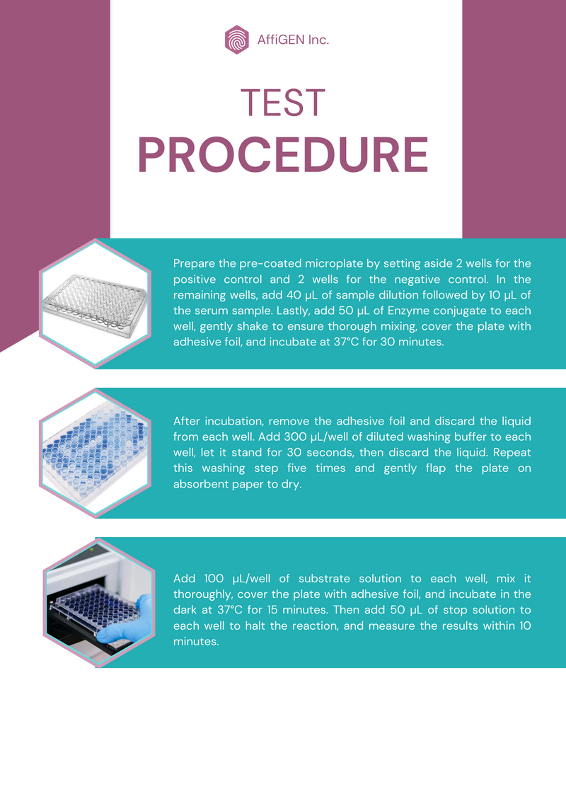
Test Procedure:
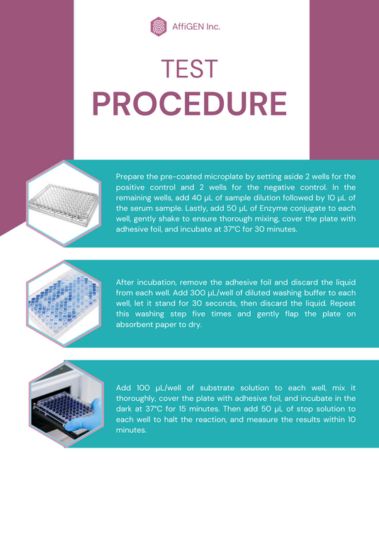
- Prepare the pre-coated microplate by setting aside 2 wells for the positive control and 2 wells for the negative control. In the remaining wells, add 40 µL of sample dilution followed by 10 µL of the serum sample. Lastly, add 50 µL of Enzyme conjugate to each well, gently shake to ensure thorough mixing, cover the plate with adhesive foil, and incubate at 37°C for 30 minutes.
- After incubation, remove the adhesive foil and discard the liquid from each well. Add 300 µL/well of diluted washing buffer to each well, let it stand for 30 seconds, then discard the liquid. Repeat this washing step five times and gently flap the plate on absorbent paper to dry.
- Add 100 µL/well of substrate solution to each well, mix it thoroughly, cover the plate with adhesive foil, and incubate in the dark at 37°C for 15 minutes. Then add 50 µL of stop solution to each well to halt the reaction, and measure the results within 10 minutes.
Result Interpretation:
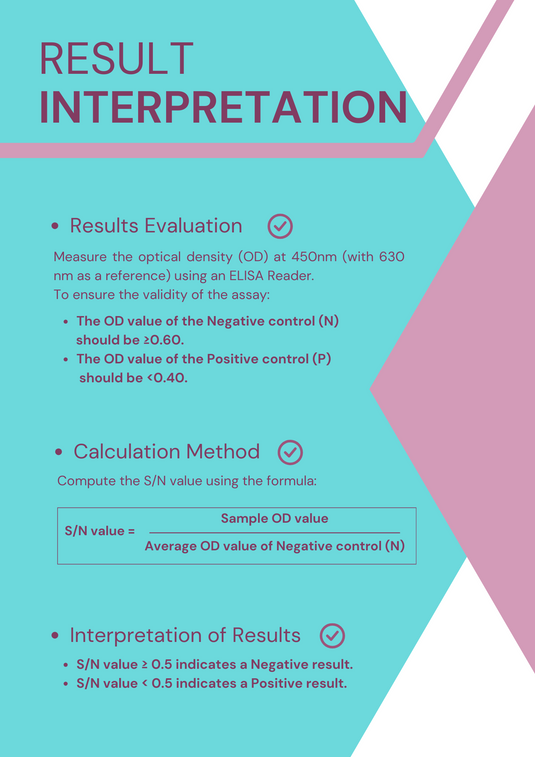
Results Evaluation:
Measure the optical density (OD) at 450nm (with 630 nm as a reference) using an ELISA Reader
To ensure the validity of the assay:
- The OD value of the Negative control (N) should be ≥0.60.
- The OD value of the Positive control (P) should be <0.40.
Calculation Method:
Compute the S/N value using the formula:

Interpretation of Results:
- S/N value ≥ 0.5 indicates a Negative result.
- S/N value < 0.5 indicates a Positive result.
Precautions & Warnings:
- 1) Read the Manual Carefully: Prior to use, thoroughly review the provided manual for detailed instructions and guidelines.
- Check Expiry Dates: Do not use reagents past their expiration dates, and avoid mixing reagents from different lots to maintain assay reliability.
- Safe Disposal: Dispose of experimental waste appropriately. Sterilize waste using high-pressure steam at 121°C for 30 minutes or treat it with a 5.0g/L sodium hypochlorite disinfectant for 30 minutes before disposal.
- MicroWell Plate Handling: If a MicroWell plate has been stored in a refrigerated environment, allow it to equilibrate to room temperature and dry before opening. Unused MicroWell plates should be returned to a dry foil bag and sealed at 4°C for storage. Liquid reagents should be capped and stored at 2-8°C in the dark, separate from other kit components.
- Use of Micropipettor: Employ a Micropipettor to accurately add samples and reagents to minimize errors. Periodically verify the accuracy of the Micropipettor.
- Washing Buffer Handling: When adding washing buffer, ensure wells are filled adequately but without overflow. This prevents the appearance of free enzyme at the well's mouth and avoids cross-contamination between wells.
- Safety with Stop Solution: Be cautious when handling the stop solution, as it is corrosive. In case of skin or clothing contact, promptly rinse with a large amount of water to mitigate any potential harm.
- Once the assay is completed, it's essential to store the kit properly. Keep it stored at 2~8°C in a dark environment, ensuring it is not frozen. The kit has a shelf life of 12 months from the date of manufacture.