

Cyclospora Molecular Diagnostics & Genomic Surveillance
Molecular Diagnostics & Genomic Surveillance of Cyclospora cayetanensis in Foodborne Outbreaks
How real-time PCR, targeted sequencing and integrated molecular epidemiology improve the detection, clustering and source investigation of Cyclospora outbreaks.
Cyclospora cayetanensis is a human intestinal coccidian parasite that causes cyclosporiasis, a foodborne illness frequently associated with prolonged or recurrent watery diarrhea. Although microscopy remains useful for identifying oocysts in stool, modern outbreak investigations increasingly depend on real-time PCR, targeted next-generation sequencing and integrated molecular epidemiology.
These technologies can detect small quantities of parasite DNA, compare isolates from multiple patients and support the investigation of contaminated produce, agricultural water and production environments. Their results are most informative when interpreted together with patient interviews, food-distribution records, environmental sampling and product traceback.
What Is Cyclospora cayetanensis?
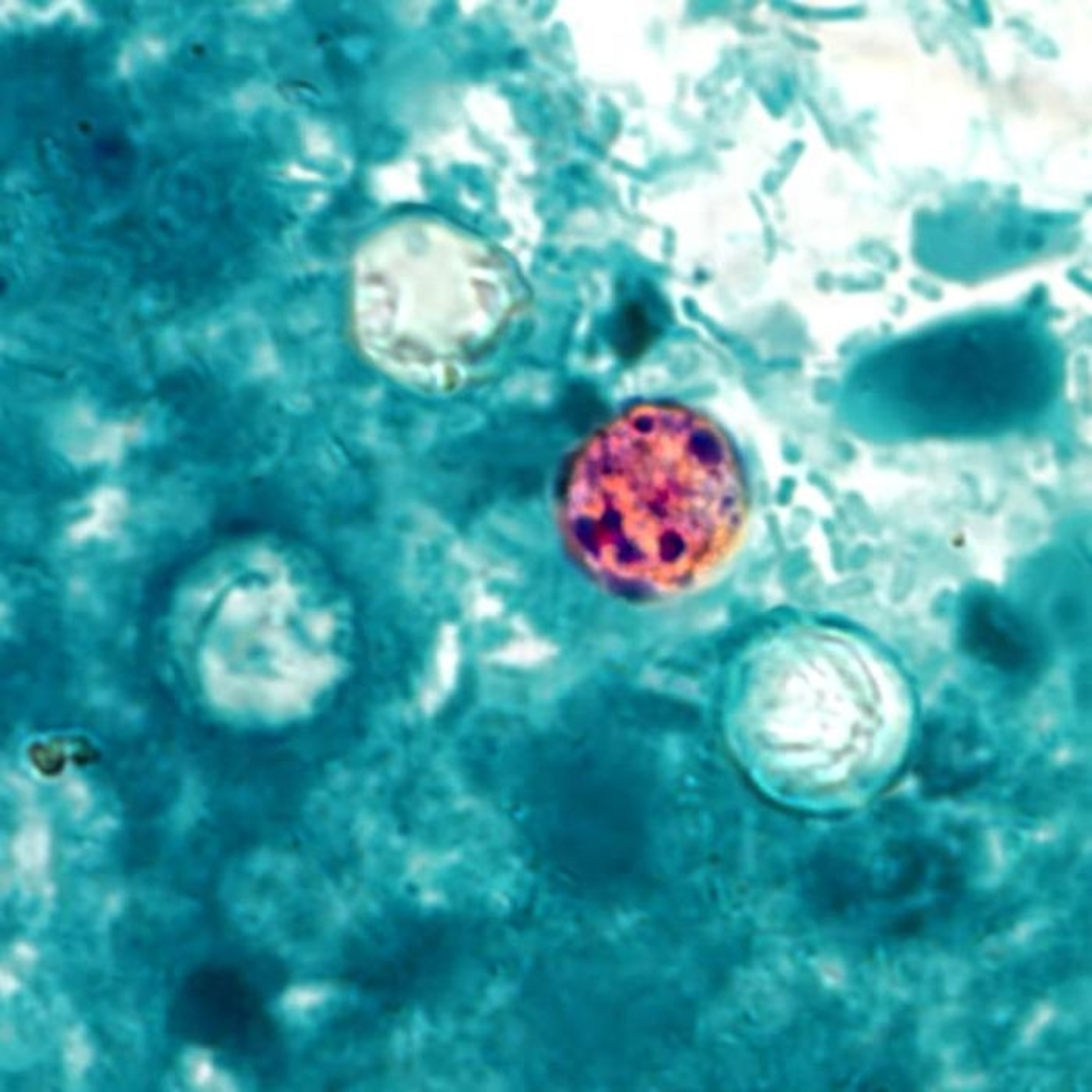
Cyclospora cayetanensis is a microscopic, single-celled parasite that infects the human small intestine. Infection occurs after a person consumes food or water contaminated with environmentally matured, or sporulated, oocysts.
Freshly excreted oocysts are not immediately infectious. They must remain under suitable environmental conditions before sporulation occurs. This biological requirement helps explain why direct person-to-person transmission is considered unlikely, while contaminated agricultural water, soil, equipment and fresh produce remain important outbreak pathways.
Human-associated parasite
Human infection is the central public-health concern, and contamination indicates a possible connection to human fecal material.
Low-level contamination
Only a small number of oocysts may be present in a food or environmental sample, making recovery and concentration critical.
No routine culture method
The organism cannot be routinely amplified by conventional microbiological culture, increasing reliance on microscopy and molecular assays.
Why Are Cyclospora Outbreaks Difficult to Investigate?
Long and variable incubation period
Symptoms commonly begin about one week after exposure. Patients may struggle to remember every herb, salad, fruit or restaurant meal consumed during the relevant period.
Short shelf life of implicated foods
Fresh produce may be consumed or discarded before investigators recognize an outbreak and identify a suspected vehicle.
Uneven contamination
Oocysts may be distributed heterogeneously. One portion of a production lot may test positive while another portion tests negative.
Simultaneous unrelated outbreaks
Multiple outbreaks can occur during the same seasonal period, making molecular differentiation and epidemiological linkage especially important.
Molecular Diagnosis of Cyclospora Infection
Microscopic examination
Traditional diagnosis is based on detecting spherical Cyclospora oocysts in stool using concentration procedures, modified acid-fast or safranin stains, wet preparations and ultraviolet fluorescence microscopy. Microscopy can be effective, but oocyst shedding may be low or intermittent, staining may be variable and identification requires trained personnel.

Conventional PCR
Conventional PCR detects parasite-specific DNA after nucleic-acid extraction. Amplified products are usually assessed by gel electrophoresis or sequencing. It can improve analytical specificity compared with morphology alone, but open-tube post-amplification processing increases handling time and contamination risk.

Real-time PCR
Real-time PCR, also called quantitative PCR or qPCR, monitors amplification through fluorescent chemistry during the reaction. It supports closed-tube detection, rapid interpretation and a reduced risk of post-amplification contamination. Cyclospora-specific assays and multiplex gastrointestinal panels may be used for clinical stool testing.

Molecular Detection of Cyclospora in Food
Testing fresh produce is more complex than testing stool because the parasite must first be removed from the food surface, concentrated and disrupted before its DNA can be amplified. The analytical sensitivity of the complete method therefore depends on more than the PCR reaction alone.
Fresh-produce qPCR workflow
The FDA Bacteriological Analytical Manual Chapter 19b describes a molecular workflow for detecting C. cayetanensis in fresh produce. The method includes produce washing or elution, concentration, nucleic-acid extraction, inhibition assessment, species-specific real-time PCR and evaluation of positive, negative and internal controls.
Agricultural-water testing
Agricultural water may introduce oocysts during irrigation, pesticide application, washing or post-harvest handling. Large-volume concentration methods such as dead-end ultrafiltration can improve the probability of recovering low-level contamination before molecular detection.
Soil and environmental matrices
Soil, wastewater, sediment and facility swabs may contain substantial amounts of PCR-inhibitory material. Reliable analysis requires matrix-specific concentration, purification and inhibition-control procedures.
| Sample matrix | Main pre-analytical challenge | Typical molecular approach | Critical control |
|---|---|---|---|
| Clinical stool | Intermittent shedding and PCR inhibitors | Target-specific qPCR or multiplex gastrointestinal panel | Internal amplification control |
| Fresh produce | Low, uneven surface contamination | Produce wash, concentration, extraction and qPCR | Process and extraction controls |
| Agricultural water | Very low concentration in a large volume | Ultrafiltration followed by extraction and qPCR | Recovery/process control |
| Soil or sediment | Strong humic and matrix inhibition | Matrix-optimized purification and qPCR | Inhibition assessment |
What Does a Positive Food PCR Result Mean?
A positive qPCR result supports
- Detection of Cyclospora-specific DNA in the tested portion.
- Evidence that the sample should receive further epidemiological or regulatory attention.
- Comparison with other laboratory, traceback and exposure findings.
It does not independently prove
- That intact, viable or infectious oocysts were present.
- That every unit from the production lot was contaminated.
- That the tested product caused the investigated illnesses.
A negative result also does not completely exclude contamination. Sparse oocyst numbers, heterogeneous distribution, incomplete recovery, inefficient lysis, DNA degradation or PCR inhibition can all reduce detection probability.
Genomic Surveillance of Cyclospora cayetanensis
Genomic surveillance compares parasite sequence information from multiple patients, foods or environmental samples. Its purpose is to determine whether specimens are genetically similar enough to support inclusion in the same epidemiological cluster.
Multilocus sequence typing
Multilocus sequence typing examines several selected nuclear loci. Analyzing multiple markers improves discrimination compared with a single locus and may reveal geographically or epidemiologically informative sequence profiles.
Targeted amplicon deep sequencing
Targeted amplicon deep sequencing amplifies selected genomic regions and sequences them at high depth. Compared with conventional Sanger sequencing, deep sequencing can detect multiple sequence variants within one specimen, an important advantage for genetically heterogeneous infections.
Expanded nuclear marker panels
Larger targeted panels can increase genomic coverage and may improve discrimination between unrelated infections. The resulting cluster assignments still depend on marker selection, sequence quality, missing data and the bioinformatic algorithm used for comparison.
Mitochondrial genome sequencing
Mitochondrial DNA is attractive for detection and typing because it may be present in multiple copies. Targeted enrichment and next-generation sequencing can recover mitochondrial sequence information from clinical or produce samples containing limited parasite material. Combining mitochondrial and nuclear markers can provide stronger discrimination than either data type alone.
Integrated Molecular Epidemiology
Genotyping provides its greatest public-health value when combined with exposure histories, product testing, distribution records and traceback information. Molecular similarity supports an outbreak hypothesis; it does not replace epidemiology.
| Investigation stage | Main objective | Typical methods |
|---|---|---|
| Clinical case detection | Confirm cyclosporiasis | Microscopy, qPCR or multiplex gastrointestinal PCR |
| Case reporting | Recognize temporal and geographic clustering | Regional and national surveillance systems |
| Molecular characterization | Compare parasite profiles | Targeted amplicon deep sequencing and multilocus analysis |
| Epidemiological investigation | Identify shared exposures | Patient interviews and standardized food-history questionnaires |
| Product testing | Detect parasite DNA in suspected food | Surface elution, concentration, extraction and qPCR |
| Environmental testing | Assess water, soil or facility contamination | Filtration, matrix-specific extraction and qPCR |
| Traceback | Identify common suppliers, distributors or farms | Purchasing and distribution records |
| Public-health action | Reduce additional exposure | Recall, advisory, sanitation review or targeted controls |
Molecular clusters can help investigators:
- Separate simultaneous Cyclospora outbreaks.
- Prioritize patients for detailed food-exposure interviews.
- Identify genetically related cases across several regions.
- Evaluate whether clinical and food-derived sequences are compatible.
- Support, refine or challenge a suspected traceback pathway.
Analytical Challenges in Cyclospora Testing
Oocyst recovery
Oocysts can adhere to irregular plant surfaces, become trapped in leaf structures or remain in discarded wash fractions. Recovery varies with matrix, sample mass, wash buffer and mixing conditions.
Oocyst disruption
The resistant oocyst wall can limit DNA yield. Validated workflows may use bead beating, freeze-thaw cycles or specialized lysis chemistry.
PCR inhibition
Plant polyphenols, polysaccharides, humic compounds and processing residues may inhibit polymerase activity. Dilution reduces inhibition but can also lower target DNA concentration.
Contamination control
High analytical sensitivity requires physical workflow separation and the routine use of no-template, extraction, positive and internal controls.
What Are the Common Food Sources of Cyclospora?
Cyclospora outbreaks have often been investigated in relation to fresh produce eaten raw and distributed through complex supply chains. Commonly investigated categories include:
Fresh herbs
Cilantro, basil, parsley and similar products may be exposed through contaminated water, soil or handling.
Leafy greens
Bagged salads, lettuce mixtures and other ready-to-eat greens are frequent targets of traceback investigations.
Berries and raw vegetables
Raspberries, vegetable trays and mixed fresh-produce products have been associated with past investigations.
Detection of Cyclospora DNA on produce suggests possible contact with human fecal contamination, directly or through contaminated water, equipment, workers or environmental sources.
Cyclospora Symptoms, Diagnosis and Treatment
Cyclospora symptoms
The characteristic symptom is frequent watery diarrhea. Other Cyclospora symptoms can include abdominal cramping, bloating, increased gas, nausea, loss of appetite, weight loss, fatigue and low-grade fever. Untreated illness may persist for weeks and can improve before returning.
Cyclospora cayetanensis treatment
Trimethoprim-sulfamethoxazole, commonly abbreviated TMP-SMX, is the standard treatment of choice for cyclosporiasis. Treatment decisions should be made by a qualified healthcare professional, especially for children, pregnant patients, immunocompromised individuals and patients with sulfonamide allergy.
Molecular genotyping is mainly used for public-health surveillance and outbreak investigation. It does not routinely determine antimicrobial susceptibility or select an individual patient’s treatment.
Future Directions for Cyclospora Surveillance
Viability-specific assays
Future methods may combine molecular amplification with viability dyes, RNA targets, metabolic markers or infectivity models to improve interpretation of whether detected oocysts remain biologically active.
Portable molecular detection
Isothermal methods such as loop-mediated isothermal amplification may support faster testing outside centralized laboratories after suitable matrix validation.
International data harmonization
Standardized markers, nomenclature, quality thresholds and interoperable bioinformatic pipelines could improve cross-border comparison of outbreak data.
One Health integration
Connecting clinical cases with agricultural water, sanitation systems, farms, processing environments and supply-chain records will strengthen source attribution.
Conclusion
Molecular diagnostics have significantly improved the detection of Cyclospora cayetanensis in clinical specimens, fresh produce, agricultural water and environmental matrices. Real-time PCR provides sensitive, target-specific detection, while targeted sequencing and multilocus analysis help identify genetically related clusters.
The strongest outbreak conclusions arise when laboratory findings are integrated with epidemiological interviews, food testing, environmental sampling and traceback records. Important limitations remain: contamination may be extremely sparse, standard PCR does not prove viability and the parasite’s genetic complexity can complicate cluster interpretation.
Continued assay validation, improved sequencing panels and harmonized international data sharing will be essential for more precise genomic surveillance and faster foodborne-outbreak response.
Frequently Asked Questions
What is Cyclospora?
Where is Cyclospora found?
Is Cyclospora contagious?
How is Cyclospora diagnosed?
How does genomic surveillance identify a Cyclospora outbreak?
Can PCR determine whether Cyclospora is infectious?
How can Cyclospora be avoided?
Scientific References
- U.S. Food and Drug Administration. BAM Chapter 19b: Molecular Detection of Cyclospora cayetanensis in Fresh Produce Using Real-Time PCR .
- U.S. Food and Drug Administration. BAM Chapter 19c: Dead-End Ultrafiltration for Detection of Cyclospora cayetanensis from Agricultural Water .
- U.S. Centers for Disease Control and Prevention. DPDx: Cyclosporiasis-Laboratory Diagnosis and Parasite Biology .
- U.S. Centers for Disease Control and Prevention. Clinical Care of Cyclosporiasis .
- Guo Y, et al. Multilocus Sequence Typing Tool for Cyclospora cayetanensis . Emerging Infectious Diseases.
- Barratt J, et al. Investigation of U.S. Cyclospora cayetanensis Outbreaks and Evaluation of an Improved Genotyping System .
- Cinar HN, et al. Molecular Typing of Cyclospora cayetanensis Using Targeted Enrichment of Complete Mitochondrial Genomes and Next-Generation Sequencing .
- Ahart L, et al. Retrospective Evaluation of an Integrated Molecular-Epidemiological Approach to Cyclosporiasis Outbreak Investigations .