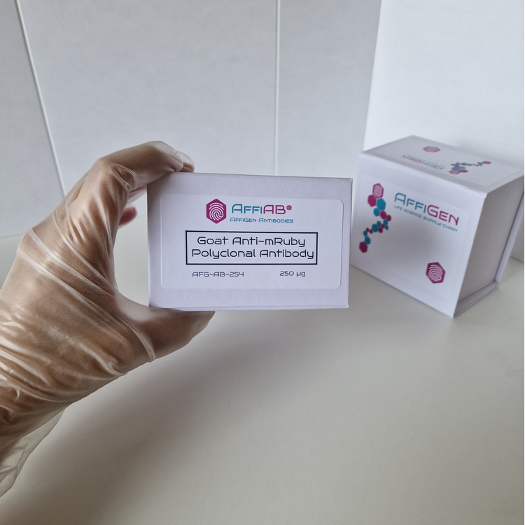
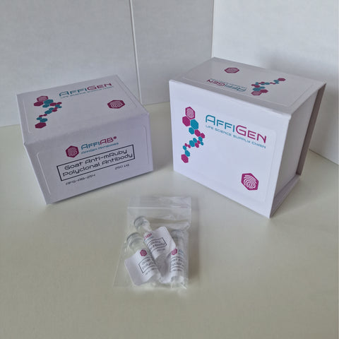
Anti-mRuby Antibody is a highly specific antibody that binds to the mRuby protein, a red fluorescent protein frequently used as a protein tag in molecular biology experiments. By binding to the mRuby protein, this fluorescent protein antibody can be used in protein localization and protein-protein interaction studies. The antibody's high specificity allows for accurate detection of the mRuby protein, making it a valuable tool for researchers conducting live cell imaging experiments.
The mRuby antibody is commonly used in combination with other molecular biology antibodies to gain insights into complex biological processes at the cellular and molecular level. These applications include monitoring protein trafficking, visualizing protein-protein interactions, and determining protein turnover rates.
Anti-mRuby Antibody is a type of antibody that specifically recognizes and binds to the fluorescent protein mRuby. The mRuby protein is a monomeric red fluorescent protein (RFP) that is widely used in various research applications, including cell imaging and protein localization studies.
Anti-mRuby Antibody is commonly used in techniques such as immunofluorescence and immunohistochemistry to detect the presence and localization of mRuby-tagged proteins within cells or tissues. The antibody is typically produced by immunizing animals, such as rabbits or mice, with purified mRuby protein to generate a polyclonal antibody that can specifically recognize mRuby.
The use of Anti-mRuby Antibody allows researchers to study the distribution and behavior of proteins tagged with mRuby, which can provide valuable insights into various cellular processes and disease mechanisms.
Anti-mRuby Antibody has a wide range of applications in various fields of biological research, including:
- Protein localization: Anti-mRuby Antibody is commonly used in immunofluorescence and immunohistochemistry techniques to detect and visualize the subcellular localization of mRuby-tagged proteins within cells or tissues.
- Protein interaction studies: By using Anti-mRuby Antibody in co-immunoprecipitation experiments, researchers can pull down mRuby-tagged proteins and identify their interacting partners.
- Protein stability assays: By monitoring the fluorescence signal of mRuby-tagged proteins over time in the presence of various treatments or conditions, researchers can assess the stability and turnover rates of these proteins.
- Live-cell imaging: The bright and stable fluorescence of mRuby allows for real-time imaging of cellular processes in live cells, and Anti-mRuby Antibody can be used to detect and track mRuby-tagged proteins in these experiments.
- High-throughput screening: Anti-mRuby Antibody can be used in high-throughput assays to screen large libraries of compounds or genetic variants for their effects on the expression or localization of mRuby-tagged proteins.
Here's a general protocol for using Anti-mRuby Antibody in an immunofluorescence experiment:
Materials:
- Anti-mRuby Antibody
- Cells expressing mRuby-tagged protein of interest
- Appropriate cell culture medium and supplements
- Phosphate-buffered saline (PBS)
- Fixative solution (e.g., 4% paraformaldehyde in PBS)
- Permeabilization buffer (e.g., 0.1% Triton X-100 in PBS)
- Blocking buffer (e.g., 1% bovine serum albumin in PBS)
- Secondary antibody conjugated to a fluorophore of choice
- Mounting medium with nuclear stain (e.g., DAPI)
Protocol:
- Grow cells expressing the mRuby-tagged protein of interest in appropriate culture medium until they reach 50-70% confluency.
- Wash cells with PBS, and fix them with the fixative solution for 10-15 minutes at room temperature.
- Rinse cells three times with PBS, and permeabilize them with the permeabilization buffer for 10 minutes at room temperature.
- Rinse cells three times with PBS, and block them with the blocking buffer for 30 minutes at room temperature.
- Dilute the Anti-mRuby Antibody in blocking buffer at an appropriate concentration, and incubate cells with the primary antibody for 1-2 hours at room temperature or overnight at 4°C.
- Rinse cells three times with PBS, and incubate them with the secondary antibody conjugated to a fluorophore of choice for 1 hour at room temperature, protected from light.
- Rinse cells three times with PBS, and counterstain the nuclei with DAPI or another appropriate nuclear stain for 5 minutes at room temperature.
- Rinse cells three times with PBS, and mount coverslips onto glass slides with the mounting medium.
- Allow the mounting medium to dry, and view the cells under a fluorescence microscope using appropriate filters for the mRuby and nuclear stains.
Note: This protocol is a general guideline, and may need to be optimized for specific cell types, fixatives, and antibodies.


