

Anti-mRuby Antibody
Detection of mRuby-Tagged Proteins in Cell Imaging and Localization Studies
Anti-mRuby is a research tool used to detect mRuby, a monomeric red fluorescent protein (RFP), and mRuby-tagged fusion proteins. It is applied in immunofluorescence (IF), immunohistochemistry (IHC), protein localization studies, and cell biology research workflows.
What Is an Antibody Anti-mRuby ?
An antibody anti-mRuby is a research-grade primary antibody that specifically recognizes and binds to the mRuby fluorescent protein. mRuby is a monomeric red fluorescent protein (mRFP) derived from the Entacmaea quadricolor sea anemone lineage. It is widely used as a genetically encoded fusion tag to visualize proteins of interest in living and fixed biological systems.
When a target protein is expressed as an mRuby fusion construct, an antibody anti-mRuby enables antibody-based detection of that tagged protein. This approach is particularly valuable when the intrinsic fluorescence of mRuby is insufficient after fixation, when signal amplification is needed, or when the experiment requires combining fluorescent protein detection with other immunostaining markers.
antibody anti-mRuby supports researchers in confirming mRuby-tagged protein expression, evaluating subcellular localization, and integrating mRuby detection into multi-channel imaging or co-staining workflows.
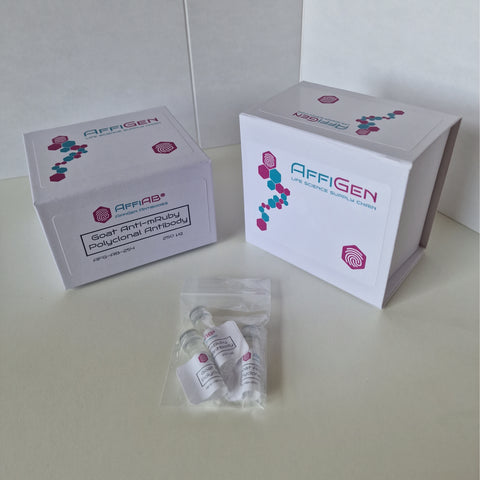
AffiAB® Goat Anti-mRuby Polyclonal Antibody
A polyclonal primary antibody raised in goat, designed for detection of mRuby fluorescent protein and mRuby-tagged fusion proteins in immunofluorescence, immunohistochemistry, and protein localization research.
View ProductWhy Use an antibody anti-mRuby?
Although mRuby is intrinsically fluorescent, antibody-based detection offers several practical advantages in research workflows. Chemical fixation can reduce or abolish the native fluorescence of mRuby, making antibody staining essential for fixed-sample analysis. Additionally, primary antibody detection followed by a fluorophore-conjugated secondary antibody provides signal amplification, improving sensitivity in samples with low mRuby expression levels.
Subcellular Protein Localization
Detect mRuby-tagged proteins within fixed cells or tissue sections to map subcellular distribution, organelle targeting, and localization dynamics.
Expression Confirmation
Verify successful expression of mRuby-tagged fusion constructs in transfected or transduced cell lines using antibody-based detection methods.
Fixed-Cell Immunofluorescence
Enable immunofluorescence staining of fixed and permeabilized samples where native mRuby fluorescence may be reduced or lost after chemical fixation.
Multi-Color Co-Staining
Combine antibody anti-mRuby detection with other primary antibodies to study co-localization, protein interactions, and spatial relationships within the same sample.
Common Applications of antibody anti-mRuby
| Application | How antibody anti-mRuby Is Used |
|---|---|
| Immunofluorescence (IF) | Detects mRuby-tagged proteins in fixed cells; supports co-localization studies with other fluorescent markers or antibody targets. |
| Immunohistochemistry (IHC) | Visualizes mRuby-tagged targets in formalin-fixed or frozen tissue sections when compatible antigen retrieval and detection systems are used. |
| Subcellular localization studies | Maps the intracellular distribution of mRuby-tagged proteins across compartments such as the nucleus, cytoplasm, mitochondria, or plasma membrane. |
| Protein–protein interaction workflows | Supports co-immunoprecipitation (co-IP) or proximity ligation assay (PLA) strategies involving mRuby-tagged bait proteins, depending on protocol compatibility. |
| Cell biology and trafficking research | Enables study of protein trafficking, turnover, secretory pathway dynamics, and expression patterns of mRuby-tagged constructs in fixed samples. |
Antibody anti-mRuby in Fluorescent Protein Research
Genetically encoded fluorescent protein tags — including GFP, mCherry, mRuby, mScarlet, and TagRFP — are foundational tools in modern cell biology. mRuby is a monomeric red fluorescent protein with excitation and emission peaks suitable for use alongside green and far-red fluorescent markers, making it well-suited for multi-color imaging experiments.
In these workflows, an antibody anti-mRuby provides an orthogonal detection strategy. Rather than relying solely on the intrinsic fluorescence of the mRuby tag, researchers can use antibody staining to amplify signal, confirm protein identity, or integrate mRuby detection into immunostaining panels that include antibodies against endogenous proteins.
This approach is especially relevant in fixed-tissue studies, high-content imaging, and experiments where signal-to-noise ratio is critical for accurate interpretation.
General Immunofluorescence Protocol Using antibody anti-mRuby
The following workflow outlines a standard immunofluorescence procedure for detecting mRuby-tagged proteins in fixed cells. All steps should be optimized for the specific cell line, fixation method, antibody concentration, and imaging system used.
Recommended Materials for Anti-mRuby Immunofluorescence
- Cells or tissue samples expressing an mRuby-tagged protein of interest
- Anti-mRuby primary antibody (e.g., AffiAB® Goat Anti-mRuby Polyclonal Antibody)
- Species-matched, fluorophore-conjugated secondary antibody
- Phosphate-buffered saline (PBS), pH 7.4
- Fixative solution (e.g., 4% paraformaldehyde in PBS)
- Permeabilization buffer (e.g., 0.1–0.3% Triton X-100 in PBS)
- Blocking buffer (e.g., 1–5% BSA or normal serum in PBS)
- Mounting medium with or without DAPI
- Fluorescence microscope with appropriate excitation and emission filter sets
Tips for Optimizing antibody anti-mRuby Staining
Titrate the primary antibody
Begin with the supplier's recommended dilution range and perform a titration series to identify the concentration that gives the best signal-to-background ratio for your sample.
Include appropriate controls
Run non-transfected cells, no-primary-antibody controls, and known mRuby-positive samples in parallel to assess specificity, background, and staining consistency.
Protect fluorophores from light
Keep secondary antibody solutions and stained samples protected from light at all times to prevent photobleaching and preserve fluorescence signal quality.
Verify spectral compatibility
Choose secondary antibody fluorophores with minimal spectral overlap with other dyes or fluorescent proteins used in the same experiment to avoid bleed-through artifacts.
FAQ: Antibody anti-mRuby
What is an antibody anti-mRuby used for?
Can antibody anti-mRuby be used for live-cell imaging?
Why use an antibody if mRuby is already fluorescent?
Which secondary antibody should be used with antibody anti-mRuby?
Is antibody anti-mRuby for research use only?
What is mRuby and how does it differ from mCherry or other red fluorescent proteins?
Explore Antibody Anti-mRubyfor Tagged Protein Detection
AffiAB® Goat Anti-mRuby Polyclonal Antibody is a research-grade primary antibody for detection of mRuby and mRuby-tagged fusion proteins. It supports immunofluorescence, immunohistochemistry, subcellular localization studies, and multi-color fluorescence microscopy workflows.
View Antibody Anti-mRuby